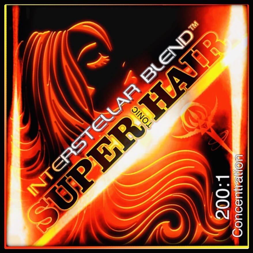
$1400 VALUE for $575! Save 59%!
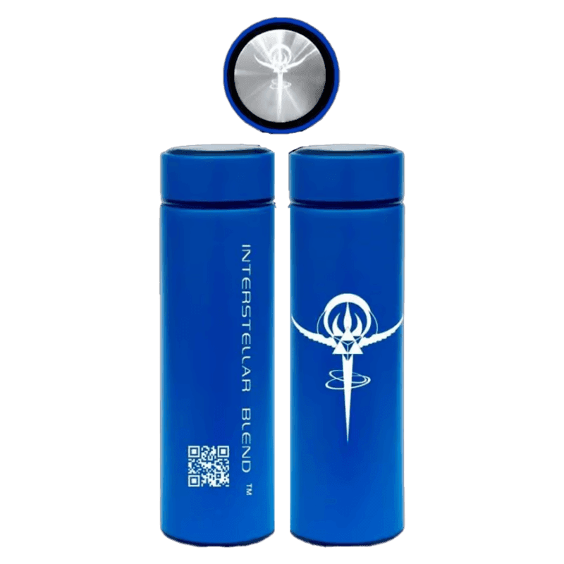
350g (50g x 7) = approx 144 1/8 tsp servings of each blend.
SUPER
hair growth
PROTOCOL
How does it work?
INSULIN blocks SHBG
SHBG blocksDHT
⬇️ LOW INSULIN =
⬆️ HIGH SHBG =
⬇️ LOW DHT =
THICK, STRONG, HEALTHY, FAST GROWING HAIR!
✨❤️✨
The OPPOSITE looks like this:
⬆️ High blood sugar =
⬇️ Low insulin sensitivity =
⬆️ High insulin secretion =
⬇️ Low SHBG =
⬆️ High DHT =
Frail, thinning hair and baldness
Low sex‐hormone binding globulin levels in young women with diffuse hair loss
We measured plasma sex-hormone binding globulin (SHBG ) and testosterone levels in a pilot study of eight women aged 21–41 years who complained of diffuse hair loss; and subsequently in a larger group of fifteen patients of a similar age range. There was a significant reduction in SHBG levels in both groups of patients when compared to controls, but testosterone values were normal.
Sex hormone‐binding globulin levels in men with androgenetic alopecia
Sex hormone binding globulin (SHBG ), plasma testosterone and saliva testosterone were measured in sixty‐four men with androgenetic alopecia and in forty males within the same age range without alopecia . There was a significant reduction in SHBG levels in bald men, compared with controls. Plasma testosterone levels were not raised in bald men, but their salivary testosterone levels were significantly higher than in controls.
Androgens play an important role in the pathogenesis of androgenetic alopecia (Ebling & Rook, 1979), but no correlation has been found between the plasma androgen level and androgenetic alopecia (Burton et al., 1979). In order to clarify this point we have studied plasma testosterone , saliva testosterone and plasma sex hormone binding globulin (SHBG ) levels in a group of males with androgenetic alopecia.
Hormonal Profile of Men with premature balding
There was a significant reduction in SHBG levels in bald men, compared with controls significantly lower SHBG values in balding men. Low SHBG is often associated with insulin resistance or hyperinsulinaemia. Another possible cause of the increased androgen effect on the dermal papilla of balding men
Results: The frequency of subnormal values in SHBG , FSH, testos-terone and epi testosterone (but not in free androgen index) was significant in the balding men. A borderline significant trend was recorded with respect to increased levels in 17OH-P and prolac- tin. Conclusions: The hormonal pattern of a substantial number of men with premature balding resembles in some respects the hormonal pattern of women with polycystic ovary syndrome.
Sex hormone-binding globulin (SHBG) production in humans has been thought to be stimulated by estrogens and thyroid hormone and inhibited by androgens. However, recent data indicate that SHBG production in vitro is stimulated by both androgens and estrogens. This study was designed to determine what other hormonal factors regulate SHBG production. Since hyperinsulinemia and hyperprolactinemia both occur in disease states in which low serum SHBG levels are found, the effects of insulin and PRL were compared to and/or studied in combination with estradiol (E2,) T4, and testosterone (T) in a human hepatoma cell line (Hep G2). Hep G2 cells were grown to near confluence in medium including 10% fetal calf serum, and then 72-h experimental incubations were carried out which used only fetal calf serum-free medium.
Compared to control incubations, both insulin (10−8 mol/L) and PRL (10−8 mol/L) decreased SHBG production from 65.0 ± 0.6 (±se) to 46.8 ± 1.1 and 46.8 ± 1.2 nmol/108 cells, respectively (P < 0.01). insulin also inhibited both E2 and T4-stimulated SHBG production. T stimulated SHBG production to the same degree as E2.
Finally, both E2 and insulin significantly increased cell number, an important consideration when expressing the effect of a hormone on SHBG production in cultured cells. We conclude that insulin and PRL inhibit SHBG production and confirm that T4, T, and E2 stimulate SHBG production in vitro. These findings suggest that insulin and PRL may be important factors in the regulation of SHBG production in vivo. (J Clin Endocrinol Metab67: 460, 1988
To correct hair loss you need to correct insulin resistance and hyperinsulinemia.
SciELO – Brazil – Low levels of sex hormone-binding globulin and hyperproinsulinemia as markers of increased pancreatic ß-cell demand in men. Low levels of sex hormone-binding globulin (SHBG) are considered to be an indirect index of hyperinsulinemia, predicting the later onset of diabetes mellitus type 2. In the insulin resistance state and in the presence of an increased pancreatic ß-cell demand (e.g. obesity) both absolute and relative increases in proinsulin secretion occur.
In the present study we investigated the correlation between SHBG and pancreatic ß-cell secretion in men with different body compositions. Eighteen young men (30.0 ± 2.4 years) with normal glucose tolerance and body mass indexes (BMI) ranging from 22.6 to 43.2 kg/m2 were submitted to an oral glucose tolerance test (75 g) and baseline and 120-min blood samples were used to determine insulin, proinsulin and C-peptide by specific immunoassays. Baseline SHBG values were significantly correlated with baseline insulin (r = -0.58, P<0.05), proinsulin (r = -0.47, P<0.05), C-peptide (r = -0.55, P<0.05) and also with proinsulin at 120 min after glucose load (r = -0.58, P<0.05). Stepwise regression analysis revealed that proinsulin values at 120 min were the strongest predictor of SHBG (r = -0.58, P<0.05).
When subjects were divided into obese (BMI >28 kg/m2, N = 8) and nonobese (BMI <FONT FACE=”Symbol”>£</FONT>25 kg/m2, N = 10) groups, significantly lower levels of SHBG were found in the obese subjects. The obese group had significantly higher baseline proinsulin, C-peptide and 120-min proinsulin and insulin levels. For the first time using a specific assay for insulin determination, a strong inverse correlation between insulinemia and SHBG levels was confirmed.
The finding of a strong negative correlation between SHBG levels and pancreatic ß-cell secretion, mainly for the 120-min post-glucose load proinsulin levels, reinforces the concept that low SHBG levels are a suitable marker of increased pancreatic ß-cell demand.
A cell-based system for screening hair growth -promoting agents
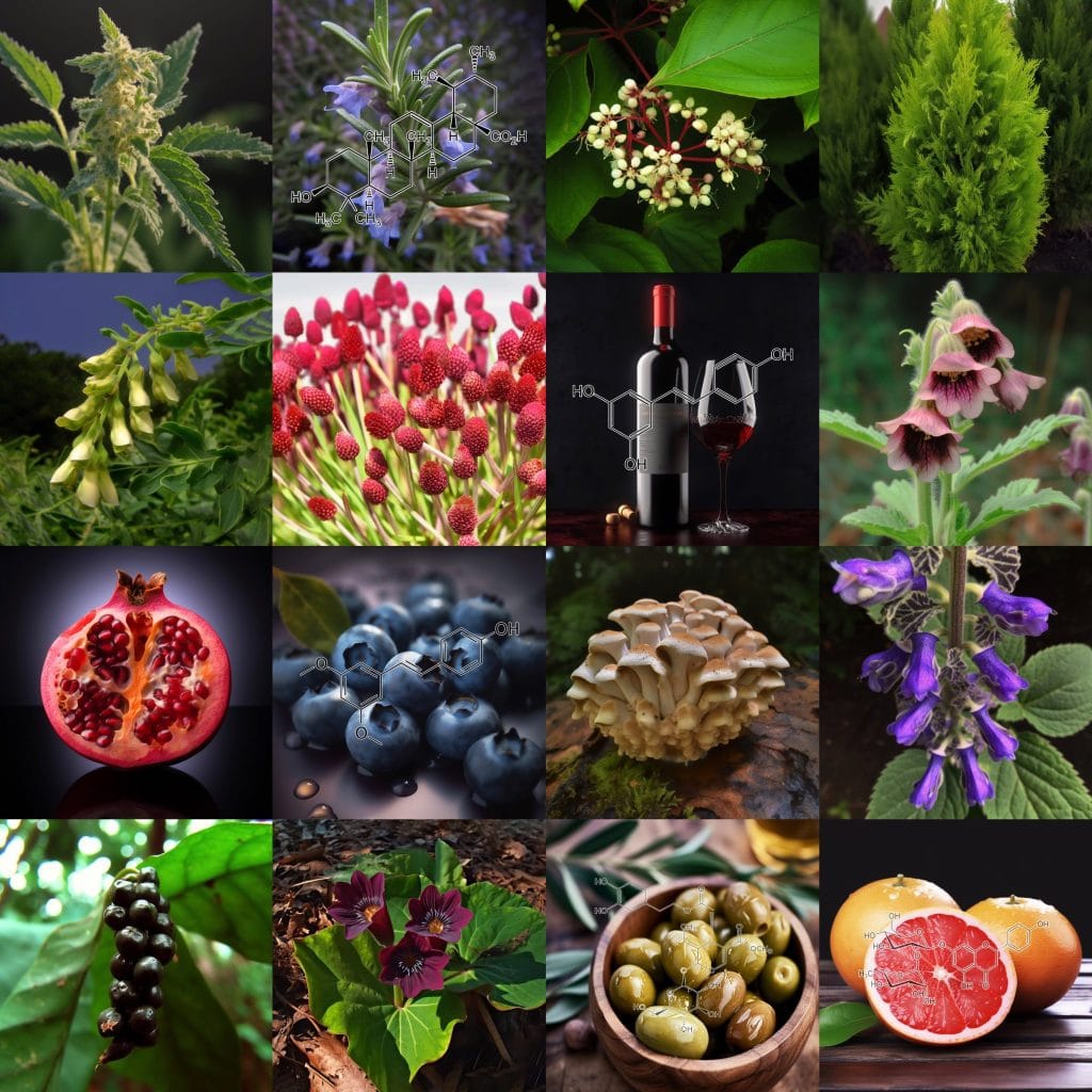
APIGENIN keeps your hair in anagen growth phase; pair with SUPER TONIC hair.
“Furthermore, apigenin stimulated the elongation of hair follicles. Taken together, these findings suggest that apigenin, which is known to have antioxidant, anti-inflammatory, and anti-tumor properties, stimulates hair growth through downregulation of the TGF-beta 1 gene.”
Androgen-inducible transforming growth factor beta (TGF-beta 1) derived from dermal papilla cells (DPCs) is a catagen inducer that mediates hair growth suppression in androgenetic alopecia (AGA). In this study, a cell-based assay system was developed to monitor TGF-beta 1 promoter activity and then used to evaluate the effects of activated TGF-beta 1 promoter in human epidermal keratinocytes (HaCaT). To accomplish this, a pMetLuc-TGF-beta 1 promoter plasmid that expresses the luciferase reporter gene in response to TGF-beta 1 promoter activity was constructed. Treatment of HaCaT with dihydrotestosterone , which is known to be a primary factor of AGA, resulted in a concentration-dependent increase in TGF-beta 1 promoter activity. However, treatment of HaCaT with the TGF-beta 1 inhibitor, curcumin, resulted in a concentration-dependant decrease in TGF-beta 1 expression.
Subsequent use of this assay system to screen TGF-beta 1 revealed that HaCaT that were treated with apigenin showed decreased levels of TGF-beta 1 expression. In addition, treatment with apigenin also significantly increased the proliferation of both SV40T-DPCs (human DPCs) and HaCaT cells. Furthermore, apigenin stimulated the elongation of hair follicles in a rat vibrissa hair follicle organ culture.
Taken together, these findings suggest that apigenin, which is known to have antioxidant, anti-inflammatory, and anti-tumor properties, stimulates hair growth through downregulation of the TGF-beta 1 gene. In addition, these results suggest that this assay system could be used to quantitatively measure TGF-beta 1 promoter activity in HaCaT, thereby facilitating the screening of agents promoting hair growth.
Mechanism of action of herbs and their active constituents used in hair loss treatment
This article discusses the mechanisms via topically applied products containing herbs and their active constituents affect the hair growth process. It was reported that the mechanisms involving (1) insulin-like growth factor-I (IGF-I), (2) vascular endothelial growth factor (VEGF), (3) epidermal growth factor (EGF), (4) fibroblast growth factor 2 (FGF-2), (5) endothelial nitric oxide synthase (eNOS), (6) Wnt/β-catenin signalling pathway, (7) prostaglandin E (PGE), (8) prostaglandin F (PGF) stimulate hair growth, whereas the mechanisms engaging (1) 5α-reductase and dihydrotestosterone (DHT), (2) transforming growth factor beta (TGF-β), (3) fibroblast growth factor 5 (FGF-5), (4) prostaglandin D2 (PGD2) inhibit hair growth. The knowledge summarized in the paper may be an inspiration to create new preparations for the treatment of hair loss.
The flavonoid luteolin inhibits niacin-induced flush
Background and purpose: Sustained release niacin effectively lowers serum cholesterol, LDL and triglycerides, while raising HDL. However, 75% of patients experience cutaneous warmth and itching known as flush, leading to discontinuation. Acetylsalicylic acid (aspirin) reduces this flush only by about 30%, presumably through decreasing prostaglandin D2 (PGD2). We investigated whether niacin-induced flush in a rat model involves PGD2 and 5-HT, and the effect of certain flavonoids.
Experimental approach: Three skin temperature measurements from each ear were recorded with an infrared pyrometer for each time point immediately before i.p. injection with either niacin or a flavonoid. The temperature was then measured every 10min for 60min.
Key results: Niacin (7.5mg per rat, equivalent to a human dose of 1750mg per 80kg) maximally increased ear temperature to 1.9±0.2 oC at 45min. Quercetin and luteolin (4.3mg per rat; 1000 mg per human), administered i.p. 45min prior to niacin, inhibited the niacin effect by 96 and 88%, respectively. Aspirin (1.22mg per rat; 325mg per human) inhibited the niacin effect by only 30%. Niacin almost doubled plasma PGD2 and 5-HT, but aspirin reduced only PGD2 by 86%. In contrast, luteolin inhibited both plasma PGD2 and 5-HT levels by 100 and 67%, respectively.
Conclusions: Niacin-induced skin temperature increase is associated with PGD2 and 5-HT elevations in rats; luteolin may be a better inhibitor of niacin-induced flush because it blocks the rise in both mediators. Luteolin inhibited PGD2 100%.
Conclusion: Taking all of this into consideration, treatment options for androgenetic alopecia should include a low cholesterol and low glycaemic index diet, improved glucose control, and fortification with magnesium. Furthermore, the current narrative does not endorse severe caloric restriction for obvious health reasons.
Widespread expression of the transcription factor, nuclear factor (erythroid-derived 2)-like 2 (NRF2 ), which maintains redox homeostasis, has recently been identified in the hair follicle (HF). Small molecule activators of NRF2 may therefore be useful in the management of HF pathologies associated with redox imbalance, ranging from HF greying and HF ageing via androgenetic alopecia and alopecia areata to chemotherapy-induced hair loss. Indeed, NRF2 activation has been shown to prevent peroxide-induced hair growth inhibition.
Multiple parameters can increase the levels of reactive oxygen species in the HF, for example melanogenesis, depilation-induced trauma, neurogenic and autoimmune inflammation, toxic drugs, environmental stressors such as UV irradiation, genetic defects and aging-associated mitochondrial dysfunction. In this review, the potential mechanisms whereby NRF2 activation could prove beneficial in treatment of redox-associated HF disorders are therefore discussed.
– Activation of the transcription factor Nrf2 plays a crucial role in protecting human organ function, specifically scalp hair follicles, against oxidative stress-induced damage.
– Nrf2 activation in human hair follicles leads to the upregulation of genes involved in phase II metabolism, reactive oxygen species clearance, the pentose phosphate pathway, and glutathione homeostasis.
– Knockdown of Nrf2 in cultured human hair follicles confirms the regulation of key Nrf2 target genes and the reduction in reactive oxygen species levels and lipid peroxidation.
– Nrf2 activation significantly reduces premature catagen (hair growth termination) and hair growth inhibition induced by oxidative stress.
– Nrf2 activation protects against oxidative stress-induced apoptosis and reduction in hair matrix proliferation in human hair follicles.
– Oxidative stress plays a significant role in hair follicle damage and graying.
– Chemotherapy can cause hair loss by inducing hair follicle dystrophy.
– Thyroid hormones and melatonin have a direct impact on hair follicle functions.
Hair loss affects men and women of all ages. Dermal papilla (DP) plays a crucial role in regulating the growth and cycling of hair follicles. Lactoferrin (LF) exhibits a wide range of biological functions, including antimicrobial activity and growth regulation. However, its effect on DP and its role in hair growth remain unknown. In this study, we found that bovine LF (bLF) promoted the proliferation of DP cells and enhanced the phosphorylation of Erk and Akt.
The bLF-mediated proliferation was significantly blocked by the Erk phosphorylation inhibitor PD98059 or the Akt phosphorylation inhibitor LY294002. Moreover, biotin-labeled bLF could bind to DP cells, and the binding was independent of lipoprotein receptor-related protein 1, a known LF receptor. Importantly, bLF stimulated hair growth in both young and aged mice. Moreover, we also found that bLF significantly induced the expression of Wnt signaling-related proteins, including Wnt3a, Wnt7a, Lef1, and β-catenin.
The bLF-mediated DP cell proliferation could be significantly reversed by the Wnt pathway inhibitor XAV939. Our findings suggest that bLF promotes hair growth in mice and stimulates proliferation of DP cells through Erk/Akt and Wnt signaling pathways. This study highlights a great potential of the use of bLF in developing drugs to treat hair loss.
💥Exercise increases insulin sensitivity (60 min cardio daily)
Any exercise that lasts longer than 60 minutes is believed to inhibit the progression of androgenic alopecia When performed routinely and consistently, the blood testosterone level will peak, then decrease in response to the body’s requirements. In addition, an extended duration of exercise will accelerate blood circulation in the scalp, increasing oxygen intake to the scalp.
This can ultimately enhance hair loss, local ischemia of hair follicles, and hypoxic conditions and prevent hair follicle atrophy.To prevent and slow the progression of alopecia, patients with normal cardiopulmonary function can be advised to engage in exercise lasting longer than 60 minutes.
350g (50g x 7) = approx 144 1/8 tsp servings of each blend.
Shelly –
Wow! I am truly amazed at how quickly my scalp stopped itching only a few days of using this product! Gavin and his crew are stellar in what they do, I am a lifetime customer. This new Super HairTonic will not leave you disappointed! Once again, Interstellar crushed it!!!! Thank you for always providing quality top notch products!
Chad –
I ordered Super tonic hair as soon as it became available. First off, the taste when mixed into coffee makes the coffee taste better. This product is one you will look forward to buying again and again. After using for a week not only did my energy levels increase, but also my hair seems to be shiny, thicker and generally easier to work with. I wish that I had this product a year ago. Would’ve saved me a lot of money buying other hair products that didn’t work well. What an extraordinary product! Thank you Interstellar!! For the quality products and customer service!!!!
Rich Ryan –
I’ve always had thin hair and after about age 40 I started getting more and more hair in my combs and brushes. I wasn’t going bald, but my hair was breaking very easily. Haircuts became optional as my hair would only grow to shoulder length before breaking off. My hair and beard felt brittle and dry, and the skin underneath my beard dried out and itched constantly unless treated with something. Also most of my beard is grey now. I can see SuperHair slowly improving all these things. There’s less hair in my comb, my hair and beard feels softer and less brittle, and my scalp and facial skin under the beard feels healthier and itches considerably less. The grey hasn’t reversed yet, but I’ve only been on the blend for a couple weeks. I’ll post an update in a year or so when the blend has had a chance to work. Once again, an incredible product from Interstellar Blends!
Tanya –
I’ve been waiting a little to post a review on this product as I try to provide as much honest and authentic detail as possible. I have never been that girl with amazing hair. I’m not saying it’s bad but it’s fine and lacks volume. Combine that with thyroid issues and it had been dry and broken in the past. I’ve been on this blend about a month and this past week I’ve started to have great hair days. (Previously I may have had one great hair day per year! My hair is fuller, has more volume and shine. I’m totally sold on this blend. This one does take a little time I think – so give it an honest go
Soma Felfoldi –
First of all, I would just like to say that Im extremely grateful to Gavin! I have been in this health field for almost half a decade and tried numerous supplements on the market but I barely got any reaction to them sadly. I already started experimenting with the dry fast thank to this site and since my Peel, Super hair and pine pollen arrived the whole dry fasting became just so easy that I feel like I never want to go back to eating. And its only 9-10 days that I have been using them! What will happen after spending some months on the protocol? Holy Moly Im excited!! After I take the blends my brain just clears out, and I am able to use the brain just like when I was in my high school years- sharp, quick math calculations, more talkative and more convincing which is very important at my work! Since mentally Im working better it seems I can do much better at my hobby which is DIY little works for homes and carpentering. I totally believe that hands skills are coordinated by the brain and my improved performance is thanks to the mental clarity.
The main reason why I got into the blends and protocol is that I have hair and skin issues since the age of 17. Now Im 27 and I dont want to shout too early but the nasty dermatitis seems to fade away finally from my face. I cant pinpoint exactly which herb does help with that but I dont even have to. I trust Gavin and the combo blends are created to work synergestically so they cannot not work! I always knew fruits and fruits skins are healthy but were never able to eat enough of them due to getting bloating. Now finally I can take the active substance from the fruits without getting bloated and this removed a huge mental burden from me that I finally get my anti oxidants. Since stressing used to increase my dermatitis and I havent had a flare up since the last 10 days!
I still have way to go on this health journey as my body temperature is lower than it should be and Im very interested in the Thermo blend and 1-2 others suggested by some members of the dry fasting group. I know my protocol is not complete yet but if its working already this well, I cant wait what will happen once I could get my hands on some more blends! Gavin, thank you for setting up such a supportive community which gives people hope and finally something which is rarely achieved: success and results!
Paula DeNote Zering –
I am a hairstylist. I have my own salon and have been doing hair for over 25 years. My hair is semi fine, breaks easy and of course I want it longer. I have been on the blends for other reason as well. I take several but added Super Tonic Hair for the sole purpose of it not falling out when I lose weight. Today my daughter was applying my color and asked me what was going on with the front of my hair. It is usually all white. I told her I was taking the blends and it’s supposed to help with grey. She laughed and said I now have dark hairs growing where there was all white a few months ago. She couldn’t believe it. Neither could I. While this is great for some, we need to keep this on the down low! Lol! I make my living on coloring white hair back. Just kidding. This stuff is amazing!
Amelia Jara –
I strongly believe that God is uncovering things like Interstellar blends to prepare our minds for the tough time we are facing. People are full of anxiety and fear that release toxins in the brain and damage it and consequently not able to think straight and make right choices. I have been practicing fasting for many years for the positive effect it does to my mind, and for weight lost . However, it was very hard to do and the pounds I lost would come back right after I resume my eating.. I started using interstellar blend in July 2019 and it’s been one of the most amazing thing I have done in my life. They are autophagy activating herbs. Auto means self and phagy means eating. Those herbs activate a system in the body that takes broken proteins ,cells that are weak and bacteria’s that are creating problems and metabolize them and feeds the body with them.. Even though you are not eating, your body is feeding you breakfast , lunch and dinner with the raw material it contains . I love all the blends but the ones that I am the most excited about are Autophagy, Super Hair, pine pollen and Niagara. Autophagy suppresses my hunger like crazy for the same reason I just stated. My body is feeding me. Isn’t that awesome. I am always calm and my stress level is very low .. Niagara’s is awesome. My libido was very low because of problems with my hormones. The first week after I started using Niagara , my intimacy with my husband increased tremendously ,sex libido and stamina increased and painful sex disappeared. Super hair combined with pine pollen are amazing. I have very dry and curly hair that never grow. Now my hair is growing and is more manageable . I also see some black hair coming out of my white hair. When I first called Gavin, he immediately sent me some samples of Thermo, nebula,trinity, peel, spice , Seven Sages ,Shilajit and matcha green tea. The first week using them revolutionized my entire body and soul. I was losing so much weight that my husband started to complain. I moved from size medium to small and extra small. My pants size are from youth 3 to 5 and that’s because I eat a lot when I eat because my husband doesn’t like it when I am too skinny. Almost a month after I started using the herbs I went to a resort at the beach eating 3 huge meals a day and snacking and drinking in between meals for 10 days and when I came back to my normal fasting routine with the blends , the first week my body looked like if I didn’t go on vacation. I am always with a strong sense of hope and good expectations for the future. I don’t get tired or sick. Praise God! I added ACB and Luteolin. My mind is so sharp that I can dream for a better future with the expectancy of living a long and happy life. Every week I either do 88/8 or 66/6 which means don’t eat food for 88 hours and just take the blends every 4 hours , then feed for 8 hours. Then the other 2 days I do intermitting fasting. This is life saving for me. I lose a lot of weight , my mind is sharp at work and I don’t have to go to the stress of preparing food to take to work. Now I am not worried
about gaining weight. My body only craves for healthy food and I am never tempted to eat the unhealthy food. My body is free from pain and inflammation and I am always looking forward to fast with the interstellar blends because of the sense of strength and encouragement I experience during the fast. I feel the presence of God in a strong way and I feel that I am stronger than the problems I face. I am always waiting for the next day with joy and positive expectations . Your body is your best friend that will be with you for the rest of your life and it is the vehicle that takes you to the places that bring you satisfaction and fulfillment in life. Therefore give it the best. Give it Intersteller blends. Your body will thank you and rejoice with you.
Rochelle Vitler –
I started using Auto, Thermo, Trinity, Super Hair and Pine Pollen approximately six weeks ago to support my overall health, mood and weight loss journey whilst undertaking a combination of 48-72 hour dry and water fasts.
I had already been fasting for approximately two months at this point but almost straightaway after introducing the blends the fasting became a breeze. I have the energy and inclination to exercise every day; I enjoy a big meal every two to three days and the desire to eat outside of these times is simply not there.
I have played around with the timing and combination of blends that works best for me and now believe I’ve got it just right. The best way to describe my mental state at the moment is “joyful”. I honestly feel happy, hopeful and uplifted, regardless of what stressors may be present in my life, and definitely attribute this to the blends.
Added bonus: I have new hair growth all around my hairline. I feel my hair overall is thicker and in much better condition but the hairline is particularly noticeable.
I can’t wait to try the next batch of blends I have ordered and will gradually add others. These blends really have made a big difference to my life and after seeing the benefits I wouldn’t want to be without them.
Claudia Gonzalez –
I purchased Superhair Tonic back in December. I had been water fasting (without blends) since August and managed to lose 30 lbs. However, by December I noticed I had started to shed tons of hair. Taking a shower caused severe anxiety since its when I saw the most hair fall out. Hand fulls of my long 30 inch hair were falling out. I had just come across the Facebook group and saw this blend. I ordered immediately. I took this combined with the Pine Pollen and within two weeks all hair loss had stopped. I was so amazed and thrilled.
I think the water fasting has also stunned my hair growth. I hadn’t seen much growth in the last couple of months. After a couple of weeks on the Superhair Tonic and Pine Pollen I noticed that my hair was growing again. I documented the growth and from the end of December to February my hair grew 3 inches! Not only that but I have tons of baby hair already growing in replacing all the hair I lost. I was afraid I would have to chop off my waist long hair with all the hair loss. But I am so happy to report that these wonderful blends have saved my hair and the 3 years that I have dedicated to growing and caring for it.
I also wanted to talk about Niagra. I’m 34 years old and have always had issues with irregular menstrual cycles. Early on they diagnosed me with PCOS. However, recently, with blood work they couldn’t really determine if that was the cause. I could go even 6-8 months without a period. I also started taking Niagra early in January. I was amazed that shortly after this I had a menstrual cycle, and then 34 days later another! No cramping, no pain, just a minor discomfort which I appreciated to know it was coming. I am so wowed at how fast this acted on regulating my cycle. I’ve felt great and will continue to take this.
I am taking other blends, like Trinity and Luteolin at nigh and my sleep has been wonderful. I truly recommend all the blends!
Qaz –
Ok finally I get to do a review! I discovered interstellar blends 3 months ago when I was searching about a certain herb. At first I thought this website was too good to be true but I looked at the hundreds of good reviews and also the reviews people have left on Facebook and I knew I had to give this a try. Now I am lucky to have great hair genetics in my family both sides of my family have a great head of hair and my dad who 52 still has a full head of hair. For me sadly I still have my full hair of hair but my scalp gets oily in one day which makes my hair brittle and fine. I’ve been taking the super hair tonic for only 3 weeks along with pine pollen and OMG, the greasy hair hasn’t gone completely yet but There’s a big improvement and it’s only been 3 weeks! Not to mention how much energy pine pollen gives me as its such a powerful super food. It’s best to take both of them together in warm water. Looking forward to trying your other blends Thank you so much a Gavin !!!
Nick K –
I’ve been taking the super hair blend as well as the spice & peel blends for about a month now.
Im impressed with the ingredient list, first and foremost. I’ve tried everything from pills to masks for my hair, and nothing comes close to this ingredient profile.
The flavor is amazing, similar to a herbal tea.
Mixing it with the Spice and Peel makes it that much better. I make it with hot water and drink it first thing in the morning.
Since taking it, I’ve noticed an increase in both thickness and shine. I’m in the process of growing my hair out, and this is one step I now realize I need to get my hair where I want it to be. I’m excited to see the progress continue over the next few months, pairing the blends with fasting and an antioxidant based diet.
anil badhan –
Amazing product, worth every penny! From the first tea I made, I felt amazing and gave me some goosebumps. The guys sent me sample of trinity also, which i combined both herbs together which made the experience way better and will be purchasing trinity next!
After few days using the herbs, I noticed how shiny my hair got. Every time I had a shower, my hair used to fall out, during and drying it. I’ve tried so many shampoos, conditioners, hair masks and oils to prevent the hair loss. This product is the best by far that I have used for hair loss.
I would recommend this to anyone with hair problems (one guy has already placed an order). Super happy how things are going and can’t wait to see the results at the end of the herbs. 100% returning back to buy more herbs! Thank you guys ! Cant wait to try new herbs! Life time customer !
Enough Love
Gloria Ho –
I love this blend. It is so easy to take this with any of my drinks twice daily. As of today, I have used the product for two weeks and have noticed a change in my hair and scalp. I do hard labor, so I sweat often and at the end of the night, I feel that my scalp is dry and itchy. When it does, hair falls out. I have tried numerous products and nothing works as well as this blend.
Since using the super hair formula, I’ve noticed my hair have stopped falling out. In addition, I’ve noticed that the necessity of my hair to be washed is less frequent because I do no experience scalp dryness or itchiness anymore. I know that washing my hair every day induces the falling out of hair, and since using this product, I can now wash my hair less frequently as well as seeing less hair fall out of my head. I really love that I can see the results right away and don’t have to take multiple pills a day in hopes that my hair would grow out. I’ve also noticed a bit of hair growth, and see baby hairs growing where there was an area in my hairline receding.
This formula is super easy to take. Every morning I mix it with my coffee and drink it easily and it does not leave a bad aftertaste. It really blends well with coffee. I also take it later in the day with any drink and I like that after today, I can reduce the intake amount. I’m very happy I got this blend and that it’s actually working. You come so many products that advertise online and promise you hair growth and you purchase it for ridiculous amount of money and subscribe to them, taking them over the.months without seeing results. With this super hair formula, I was able to feel and see the results almost immediately and am speechless with the end result.
I will definitely be a returning customer and continue to use this product alongside Pine Pollen which I also bought. This prevents hair loss and replaces so many products such as pills, shampoos, conditioners, and other home remedies I’ve tried. It is by far the best hair loss product I’ve ever used. Thank you to Interstellar for the research and effort that they have put into in order to create such a fantastic blend for hair. I can’t wait to try the other blends. I’ve also got the Triinity, Thermo, and Supernova blends, and they are fantastic as well and helps me with intermittent fasting! Thank you so much for creating this product and finding such a great blend of herbs! I’m excited to get my hair where I want it to be.
Best Regards! You’ll be hearing from me soon with additional purchases! Fantastic work you’ve done here!
Nirali –
First thing First, Interstellar blends are a game changer. I ordered Super Hair Tonic and have been taking it for about 3 weeks now and I can already see the difference.I have Wavy curls, and my hair were pretty well until I moved to Canada. After moving to Canada, I started experiencing grey hair, Hair-Fall, low hair volume and my hair started to break very easily. I tried so many supplements and hair care products but all in vain. I was so frustrated and stopped caring about my hair at all.
Then my friend introduced me to Interstellar Blends. I reached out to Gavin and he recommended Super Tonic Hair. I was very skeptical at first as everything i’ve tried until now had shown no result but I thought of giving it a try. The order came in just few days of ordering it. I have been taking the Super Tonic Hair for 3 weeks and I can literally see the difference in my hair. They are now getting longer, less hair-fall and I can also see new hairs growing. And its not just about the hair but after I started taking Super Tonic Hair, my whole body feels so different, Very light and always at ease. This blends are really life changing and I can’t thank Gavin enough for creating this blend. He is very knwoledgable and answers any question that I have right away. The Customer service is very SPOT-ON.
Over-all I am very happy with the result and I am really glad that I gave interstellar blends a try. I can’t wait to explore other blends and reset my body.
I have just become the lifetime customer of Interstellar Blends.
jennifer –
I have always had good hair, thick and wavy. but it doesn’t seem to grow much past shoulders. And despite having a good diet and pampering my hair with ‘good ” hair products, it would still get a bit dry and frizzy… after a month of using the Super Hair Tonic, my hair grew, I’m seeing new hair growth and its not dry. It looks thick and nourished. the best its ever looked and other people are noticing too… but its not just about my hair…. I feel better in general. This is the first blend bought. I’ve since bought Trinity, Nebula and have received samples of seven sages and super nova.. all amazing. I never want to be without these products. I love how I feel on them… sometimes I fast with them… but even when I don’t. I still feel the positive effects of the blends.
S.R. –
Where to begin…. I have fasted, both wet and dry, for years prior to coming across Interstellar Blends over a year ago. Like most, I lurked and read testimonial after testimonial wondering if there was actually truth to what I was reading. I am a great lover of science, so Gavin’s website was a treasure trove for me. There was article upon article convincing me that the testimonials I was reading could actually be true.
I reached out to Gavin and he quickly responded to my inquiry. That alone spoke volumes about his integrity. I then placed an order and received it within a couple of days. The packaging was SUPERB! It was so beautiful that I didn’t even want to open it out of fear of messing it up, but boy am I glad I did.
I was never a fan of coffee because it gave me the jitters, but I still followed the instructions. The blends tasted horrible so I quickly became grateful for the coffee that helped mask the taste. Within a few minutes, I started to feel different. There was something happening, but I wasn’t sure what or how to explain it. I was feeling an “opening” going on in my chest and head. Soon, my body began to crave the blends, nasty tasting and all!
I have already tried most blends, but I want to focus on Super Tonic Hair and Pine Pollen. As I mentioned previously, I was no stranger to fasting; and as most would know, that usually means hairloss. My hair had become so thin and brittle that I felt it was a lost cause. I began taking Super Tonic Hair and Pine Pollen, and within a month, I began to get compliments on my hair’s fullness and shine. I no longer had to choose between fasting and my hair. With Interstellar Blends, I can have it all!!
Thank you Gavin for sharing your wealth of knowledge and your products with the world. You and your whole team are positively impacting and changing lives worldwide!!!
Kevin –
After I noticed my hair thinned out really quick I decided to order super tonic hair, I was like: nothing to lose right, so why not try it out? After a week I immediately noticed my hair didn’t fall off easily anymore and I’m just a week in!
Also I noticed that beside growing back thicker and less falling out that the quality became better. Also it tastes really good! Like a light version of coffee which makes it part of my morning routine.
Can’t wait for what the coming months will bring, but I sure do now that I will order now right away another one.
Bal Purewal –
I feel very blessed and grateful that i started taking super hair tonic 4 weeks ago. To be honest – it was not my hair that was a priority for me as i have other issues to deal with. However , I purchased the combo pack of blends ( thought that was my best way forward to take all the blends as per Gavins 22:2 protocol . Like i said at the beginning – my hair was not really my main priority for now – main reason being i fell out with my hair 30 years ago when i got my first grey hair (i was 13 years old) – i knew this wa genetic as my family members also started going grey as children – by the time i was 27 years old i succumbed to dying my hair ( so next 24 years was a big commitment to dying my hair every 4 weeks) , in that time – i knew the hair dyes were no good – toxic and carcinogenic – i kept dying my hair as could not leave my house if i saw one white hair!!!! Then we had the first lockdown March 2020- and thats the ush i needed to stop dying my hair – however – i felt that the damage had already been done to my hair – it was thin – lacked lustre and i had over the years developed 2 very very thin (almost bald ) patches on my hairline (above my temples ) which i felt i had to accept as never grew back- Now back to the super tonic hair – started taking 4 weeks ago – one quarter teaspoon once a day only – and my hair seems to have thickened considerably – the bald patches do not look as bald anymore and I’m feeling so much confident with my hair and not afraid to go out . At this time of writing this review- i personally not sure if my grey hairs are reducing and my natural colour coming back ( it appears like it is) – i will write another update in couple months time . Needless to say i have now purchased a 100g bag – without batting an eyelid – i would advise anyone who cares about their hair – STOP GOING TO THE HAIR DRESSERS – DONT ALLOW CARCINIGENS IN HAIR DYES-AND HAIR PRODUCTS ANYWHERE NEAR YOUR SCALP AND HAIR – THE MONEY YOU SAVE NOT GOING TO HAIRDRESSERS WILL BE MORE THAN YOU NEED FOR THE SUPERTONIC HAIR – you will never go back – for the first time in my life – i love my hair – even the greys – i call myself the OCEAN PRINCESS
Suzanne Kici –
I have been using this product for approximately 12 months and I cannot believe how healthy my hair is. My stylist said that it is so much thicker and has more volume and is not dry like it used to be. I absolutely love this product!
Valerie Cade –
As a woman who is 53 with thinning hair due to menopause hormonal issues, Super Hair Tonic truly restored my thinning hair to the fullness I’d experienced before ~ and within on week. My mistake was only ordering the sample; I now realize ordering the full package is the key as this blend is a staple for me moving forward (along with peel and spice for every day muscle aches and pains; these are gone, trinity for focus and motivation and autophagy for controlling hunger as I continue to lose weight and not have any cravings). Super Hair Tonic exceeded my expectations!
Harry Aplin –
I’ve been using this product for the last two weeks and I’m very impressed.. it took about 5 days for me to notice a change, with the strands of my hair looking much thicker and healthy..after a couple of weeks I noticed my eye lashes got really long too! I’ve never seen them so long before!
I used to suffer from an itchy scalp before using this product but now it feels so good to be free of that, super happy with the results and will be buying more for sure, also keen to try out some of the other products!
Thankyou Gavin!
Patricia –
I’ve been taking the blends for over a year. LET ME TELL YOU, these are by far the BEST thing I stumbled upon. I have always been into alternative medicine and am never willing to take what big Pharma is pushing on me. I am a retired Registered Nurse and have pushed back against Pharma my entire career…weird right but my eyes were opened.
I have suffered with arthritis, pain, low energy but feeling of impending doom, thinning hair, and high blood pressure. Let me tell you….I tried many supplements to try and reverse these symptoms and nothing worked. Again by the Grace of God, I came across Gavin and his life saving blends. What a game changer!!
Here’s my results. By taking Peel and Spice for Arthritis, I have function back in my wrists and pain is under control. Low Energy and impending doom? GONE with NEBULA and TRINITY…I have never felt so calm and let things pass in my entire life! I use to be high strung and everything effected me. After having what I think was COVID..(never got tested) I started loosing my hair. How disturbing to me. I have been taking the hair blend and is thankfully growing back. Last but not least, EKG has brought my blood pressure into normal range. At every annual physical, my blood pressure was so high sometimes they didn’t want me to leave the clinic. This is a game changer for me. So happy I’m not in the critical zone anymore.
I could go on and on about the blends. Purge, Glucose Blocker, Anti Adipogenic, Thermo, and Autophagy for loosing weight…OFF THE CHARTS. I lost 10 pounds and have kept it off for over a year.
This is getting long so just try them….you’ll never go back! Thank You Gavin so much for your life saving and beautiful blends. They are filled with Love.
Kristina –
I have been taking super hair formula for 2 months now. I bought it to mainly help with gray hairs. I am 33 but started getting grays in my late 20’s. My hair has also been thinning and not growing past a particular length. I did not notice results the first 2 weeks, but kept taking the blend since I had it I figured I might as well finish it off. In the 3rd week, I noticed this bump/wart I had at the nape of my hairline disappeared. It had been there for as long as I can remember. I was shocked to run my finger where it used to be and feel smooth skin. I felt encouraged.
I have been wearing tape-in extensions for about 2 years to give myself the appearance of longer, thicker hair. 6 weeks in of taking the blend, I had a routine hair appointment to remove, clean my extensions, re-install them, and get my roots colored. As my hair stylist was removing them, she commented on how much new growth she was seeing. When she finally removed them all, we were both shocked to see my actual hair had grown as long as the extensions!!!! This has never happened before! My hair was also so much thicker. She only installed half as many as I used to get, and for my next appointment in a couple weeks, I am going to remove them altogether.
I have not yet noticed an improvement with the grays, but I think I will be able to analyze results better once I take my extensions out and take a break from hair coloring. I think it may just take longer to see that change, but I feel encouraged because of the amazing results in length and thickness. I will update after a few more months on the improvement of grays! This is a wonderful blend but it takes a few weeks to see the results. Do not be discouraged if you don’t see a change right away! Stick with it and be consistent.
Diana Ricco –
Super Tonic Hair is a life saver!!! I was starting to lose my hair in clumps after I had covid. Not a single remedy worked to stop the hair loss, let alone grow it back. I was super thinned out and I couldnt even pass a comb through my hair without losing a bunch of it. Enter this blend!
Within a month of taking it, i was sprouting fresh new hairs all over my head. My existing hair felt stronger and less brittle, AND i havent seen any random strands of grey hair since I started taking this blend. I am a repeat customer already and its been about a year now. My hair is thicker than before. It is stronger than before. It feels and looks way healthier than before.
Even my hairdressers are shocked by the drastic change in thickness and hair growth that theyve all been asking me for the name of this blend. They tell me they have NEVER seen anything like the results theyve seen on me, and they see people’s hair every day. It is truly life changing. It brought me from being stressed and depressed to being confident agian. I dont think I will ever not buy this blend for as long as I live. Thank you, Gavin.
Konstantin K –
One month ago I placed my first order and boy was I not disappointed! First of all, I can feel all of them, each has a very pronounced effect. Supernova makes me super focused and alert, gainz significantly increases my muscle mass and strength, the rest just make me feel better in general. I’ve been experimenting with all sorts of different supplements and practices over the last 2-3 years but I have never felt such prominent effects.
Although, in this review I’d like to particularly highlight Super Hair. I’ve been dealing with slowly receding hairline since I was 20. I started fighting it right when it started, doing all sorts of things from natural toos like microneedling and rosemary oil to more kind of pahrma things like minoxidil. Last few years even though I’ve been doing a lot, the receding hairline started to win the battle. I’ve been seriously thinking about doing hair transplant in the next couple of months.
Hard to put in words how amazed I was with the results I got from Super Hair. Never have I ever seen such a FAST and PRONOUNCED effect. One week in I could see new hairs growing and old ones getting thicker. No other hair solution or compound works that fast. Minoxidil takes around 8 weeks to see the first tiny results. Hats off to Gavin for creating such a powerful and natural solution. 👏👏👏
Lori Cramer –
I have been using this blend a little over a month, so I can’t give a full review as yet but I am liking what I see. Fuller thicker hair ..i definitely see a difference from before. So im pretty excited to keep using and see more results. I am definitely excited to try other blends.
Jennifer W –
It’s funny how you get used to feeling a certain way so that’s just what you accept of yourself. Or you aren’t aware that something is wrong with you until you feel better. I didn’t realize how exhausted I get during the day. I didn’t realize how low energy I am. I just go through my day and anxiously await a proper time to go to bed. Or…. what’s it feel like not to have a headache? I don’t get migraine headaches, it’s just from not sleeping. I can expect a headache at least four times a week. Stomach aches from poor digestion are awful too. Just overall feeling good… really? Yes!!! Yes!!! Yesssss!!!!
Interstellar Blends to the rescue. So I have several of the blends in sample size. I’ve actually had them quite a while and I would take some here, take some there. I really got serious about it this past week and I have taken them at least twice daily and sometimes three times daily. WOW is all I can say. The blends certainly have a way of pointing out just how poorly I felt by making me feel amazing!!
I have been taking a concoction of plush, nebula, super hair, spice, niagra, super nova, and several more. The first thing I noticed was that I can actually breathe through my nose. I have absolutely no congestion. I suffer with seasonal allergies and allergens from indoors. I work in an over 100 year old building and there’s stuff everywhere in there. I have zero congestion and zero allergy symptoms.
Energy? Ha!!! I’m freaking annoying because I have so much energy. Sleep and blends!! No more headaches. Not a single one this week. An overall mellow mood and I’m nice. I’m so, so nice now!!! LOL Digestion issues gone!! When I take steps to feel better I don’t want to put the crap in my body so I’m eating a better diet too. I’m fasting and taking the blends with coffee or tea.
The blends go above and beyond what they’re intended to help you with. All this and I haven’t even tried the blends for sleep. Since I’m headed into the weekend I will give that a shot…just so I can adjust the amount so I will get up in the morning for work when Monday rolls around.
The Interstellar Blend website is so amazing. I love the updated “choose your own symptom” scrolling technology provided. It makes choosing blends so convenient.
Thank you Gavin and Interstellar Blends. I haven’t felt this good in a very long time…. Possibly ever!!
Deb Kroon (Alana) –
I just went back to my hairdresser yesterday after months of not going. I had gotten in touch with her after my pyramid was burned to the ground and told her of my hair loss due to the anxiety and stress. She just couldn’t believe the new hair growth due to the hair blends, Super Tonic Hair and Apigenin. She was absolutely amazed. She didn’t even get my hair done before asking me for your card. Thank you just doesn’t seem like enough Gavin, you are an absolute saint who is helping so, so, so many people. I absolutely panicked while I was losing my hair. My hair is down past my butt and is a very important aspect of who I am. I am delighted at the come back. More people need to introduce their hairdressers to the blends. Men and women alike. I love them all but HAIR BLENDS ALL THE WAY FOR ME!!